

Reliable care through "team medicine",
"state-of-the-art equipment",
and "specialized expertise"
Feature 1
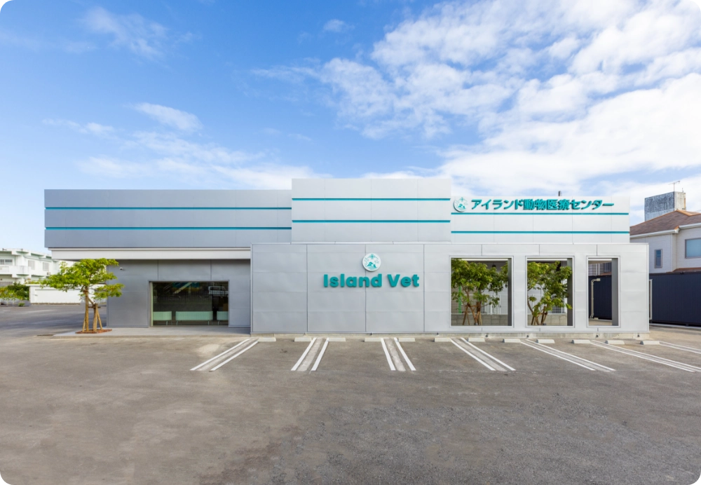
Island Vet leverages the know-how cultivated at our group hospital, Chubu Vet and is committed to providing the best possible veterinary care as a central hospital on the island.
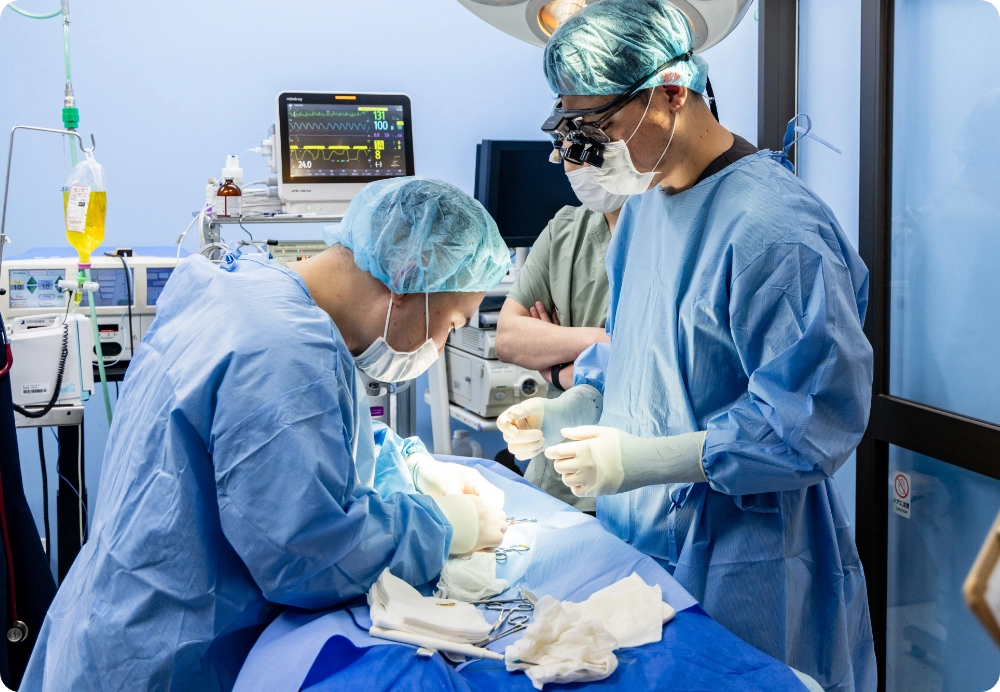
Team medicine
Together with Chubu Vet, our group comprises more than 30 medical staff. As a year-round regional provider, our team is dedicated to giving each animal our full attention. We have a culture of strong teamwork where staff collectively build and maintain collaborative care. Each veterinarian has their own areas of expertise, and case discussions among specialists improve overall care quality and provide greater reassurance and satisfaction for our patients and their owners.
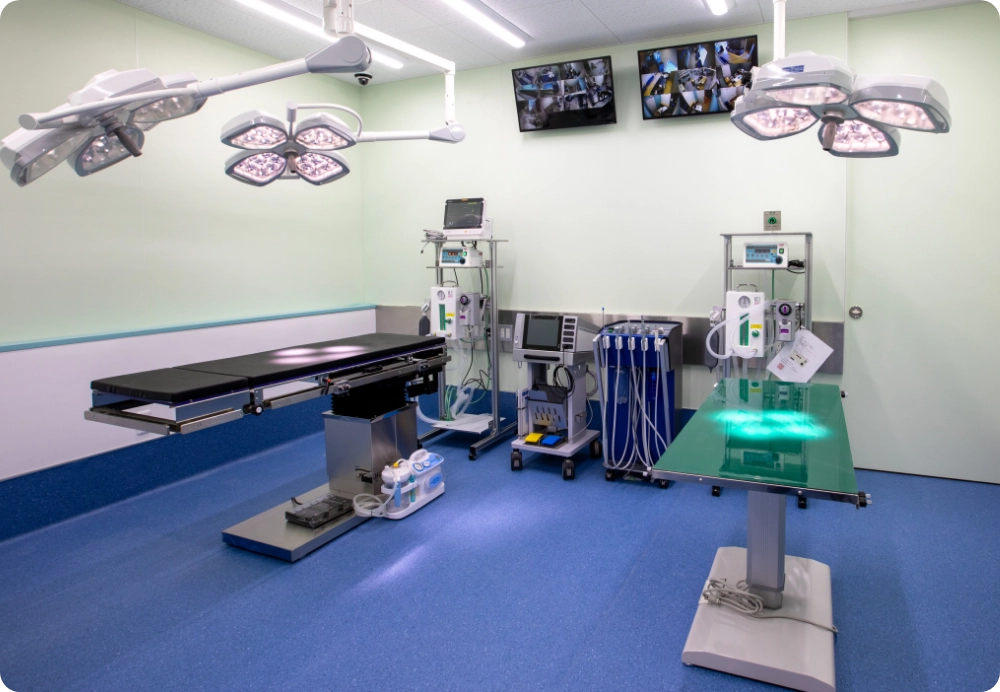
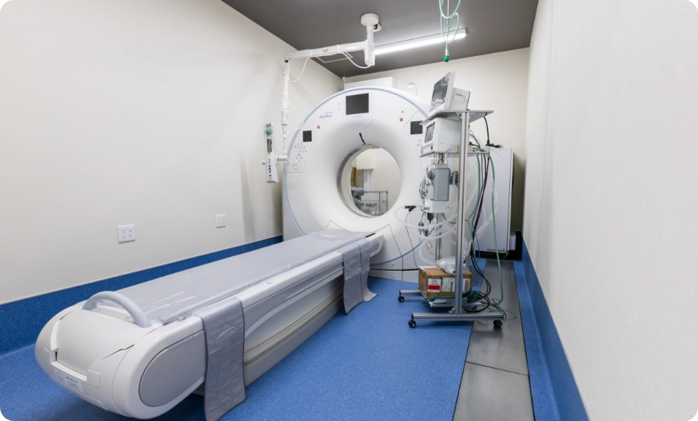
State-of-the-art equipment
We actively introduce advanced veterinary equipment that is rare in Okinawa. Our goal is to minimize the number of patients we cannot help and to contribute to the region and society. In particular, minimally invasive laparoscopic procedures are uncommon in the prefecture, and before our CT installation there were no CT scanners north of Urasoe. By installing CT in Ishikawa, Uruma, we provide broader regional access and meaningful social benefits.
Specialized expertise
There are few veterinarians with specialist knowledge in the prefecture, and appropriate treatment is sometimes unavailable. We invite multiple specialists from mainland Japan to provide consultations and care. Our system enables second-tier and specialist-level examinations and treatments; even when a specialist is not physically present, our veterinarians can consult with them to ensure appropriate care.
Introducing laparoscopic surgery
Feature 2
Smaller incisions, faster recovery.
“Keyhole surgery” that balances safety and reliability.
“Keyhole surgery” that balances safety and reliability.
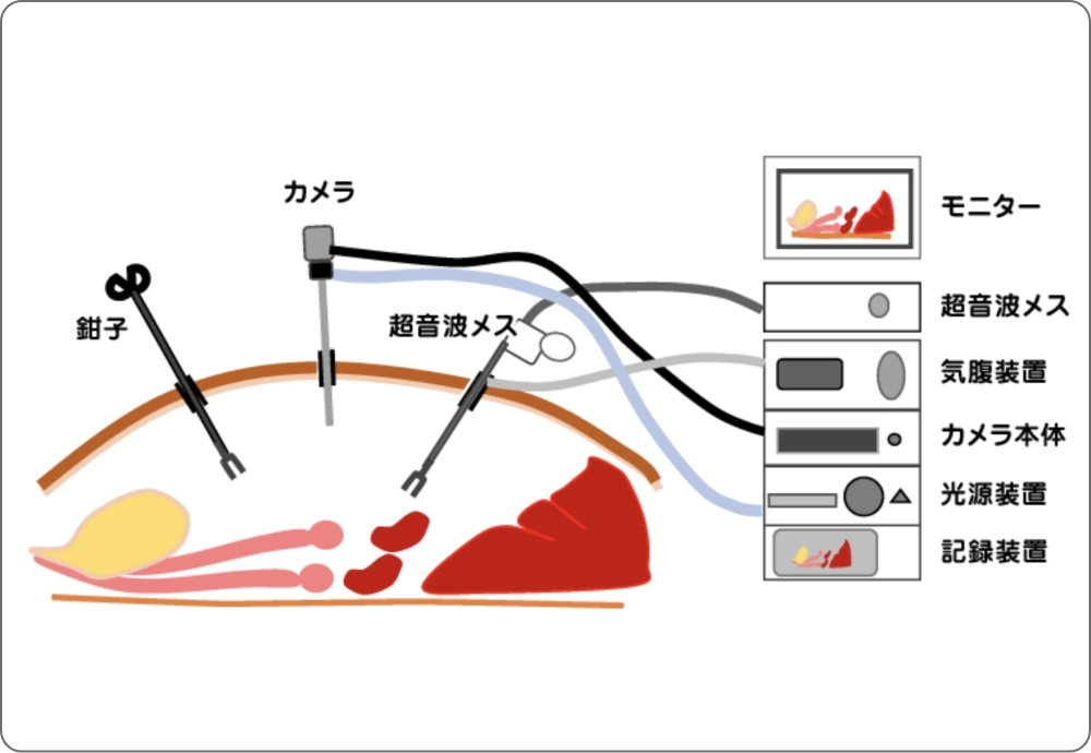
Laparoscopic surgery is performed by inserting a camera and instruments into the abdomen and operating while viewing a monitor. Incisions are small (3–10 mm), and postoperative pain is typically less than with open surgery. It reduces the burden on the animal and is a safer, gentler option.
Typical procedures suitable for this approach
- Spay surgery (ovariectomy: OVE / ovariohysterectomy: OVH)
- Preventive gastropexy (prevention of gastric dilatation-volvulus [GDV] in large dogs)
- Removal of cryptorchid testes (abdominal or inguinal)
- Biopsies of liver, kidney and other organs
- Assistance with bladder stone retrieval, evaluation of gallbladder or spleen (case dependent)
Benefits of laparoscopic surgery
- Smaller incisions: surgery can often be performed through two or three ports less than 1 cm each.
- Less pain / faster recovery: reduced tissue damage leads to quicker postoperative recovery.
- Enhanced visibility: camera magnification helps identify small vessels and organs, improving bleeding control.
Other advantages
- Fewer wound complications: reduced tension on sutures lowers risk of infection or dehiscence.
- No need to exteriorize organs as in open surgery, reducing pain from ligament stretching.
- No direct exposure of organs to air, lowering the risk of adhesions.
Drawbacks of laparoscopic surgery
- Requires advanced skills: specialized instrument handling and techniques are needed and differ greatly from conventional procedures.
- Not suitable for all cases: severe inflammation, extensive bleeding, very large masses, or dense adhesions may be safer to treat with open surgery.
- Requires specialized equipment and experience: availability may be limited.
Other considerations
- Insufflation with CO₂ may temporarily affect respiration and circulation.
- Costs are generally higher than conventional surgery.
- If unexpected complications arise during surgery, conversion to open surgery may be necessary.
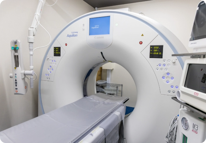
What is animal CT scanning?
Feature 3
See the body in three dimensions.
High-precision imaging to detect hidden disease early.
High-precision imaging to detect hidden disease early.
CT uses X-rays from 360° around the body and computer reconstruction to create images that detect early disease. Widely used in human medicine, we aim to make CT scanning as familiar and accessible for animals as X-ray and ultrasound.
About CT scans
Point 1
Detects very small lesions
Detects abnormalities that are easy to miss on routine tests — tumors, nasal cavity lesions, small pulmonary lesions, fractures, and more.
Point 2
Fast and broadly useful
Scanning takes only a few minutes and supports diagnosis across many fields, including tumors, neurology, trauma, and dentistry.
Point 3
Regional contribution of CT availability
CT scanners are scarce in Okinawa, and previously none were available north of Urasoe. Installing CT in Ishikawa, Uruma improves access for central areas and communities north of Urasoe, helping more animals receive timely care.
CT scan features
- Can image areas difficult to evaluate with radiographs or ultrasound.
- Provides an overall view including complex structures and small organs such as lymph nodes.
- Shows relationships between lesions and surrounding organs or vessels.
- Helps owners visualize the condition in three dimensions.
- CT can also be used for health screening.
Conditions where CT is useful
〈 Head 〉
Hydrocephalus / otitis externa / otitis media / dental disease / tumors (brain, nasal, oral, orbital) / fractures / brain disorders / altered consciousness / facial swelling
〈 Thorax 〉
Tumors (lung & metastatic lung lesions) / lung lobe torsion / tracheal collapse / fractures
〈 Spine & spinal cord 〉
(Examples include spinal trauma, intervertebral disc disease, vertebral fractures and other spinal lesions)
〈 Abdomen 〉
Tumors (liver, kidney, spleen, pancreas, adrenal, bladder, intestines, lymph nodes) / portosystemic shunt / stones (kidney, bladder, ureter) / intestinal obstruction / intussusception / foreign bodies / fractures